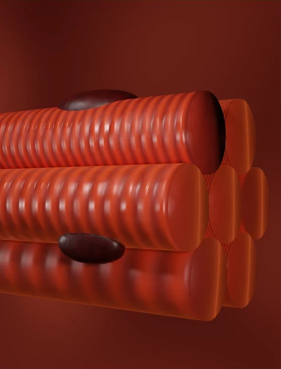
Muscles are an integral part of the human body, allowing us to move, stabilize, control bodily functions, and generate heat. There are over 600 muscles in the body, comprising about 40% of our total body weight. Understanding the anatomy of muscles can help us better comprehend how they work to enable body movements and functions. This guide will provide a comprehensive overview of skeletal muscle anatomy and physiology.
Skeletal Muscle Structure
Our muscles are attached to bones by tough cables called tendons. This lets them tug on the bones and move our bodies. Without muscles hooked up to skeletons, we would just be puddles of jelly!
Each muscle contains thousands of long, stringy muscle cells bundled up like telephone wires. These cells are stuffed with even smaller strings named myofibrils - made up of two proteins, actin and myosin. These proteins slide across each other and make the cells squeeze, shortening the muscle.
The muscle cells are bundled into cable-like groups called fascicles, wrapped in a thin layer of tissue. A bigger cable, the epimysium, binds all the fascicles together into one powerful muscle.
The muscles also have their own blood supply and nerves woven throughout them. The nerves allow the brain to activate the muscle cells in a coordinated way - telling them when to shorten and squeeze! So the muscles tug on our skeletons, powered by actin and myosin proteins sliding around inside the cells, all under signals from the nerves. This is what allows us to move!
Types of Skeletal Muscle
There are three types of skeletal muscles in the body, classified by their structure and pattern of use:
- Smooth Muscles: Found in hollow internal organs like stomach, bladder, intestines. Long, thin, spindle-shaped cells. Contract slowly but sustain contractions longer. Involuntary control.
- Cardiac Muscles: Present only in heart. Branching pattern allows electrical signals to spread quickly. Contracts involuntarily. Powers heartbeats.
- Skeletal Muscles: Attach to bones by tendons. Striated pattern. Rapid, powerful but short contractions under voluntary control. Enable body movements. Further classified into:
- Type I (Slow Oxidative): Used for sustained activities like standing, maintain posture. Highly resistant to fatigue but weak. Rely on aerobic respiration. Examples – soleus calf muscle.
- Type IIA (Fast Oxidative-Glycolytic): Used in short yet powerful bursts e.g. getting up from chair. Generate moderate strength. Use both aerobic and anaerobic respiration. Examples – abdominal muscles.
- Type IIX (Fast Glycolytic): Recruited briefly for most strenuous strength-based activities. Largest and strongest. Use mainly anaerobic glycolysis for energy. Prone to early fatigue. Examples – quadriceps, biceps.
Muscle Contraction Mechanism
Skeletal muscle contraction occurs by the sliding mechanism of actin-myosin cross-bridges when motor neurons activate the muscle:
- An action potential travels down the motor neuron into the neuromuscular junction.
- Acetylcholine is released, triggering depolarization of the muscle fiber.
- The signal spreads across the fiber surface, into transverse tubules (T-tubules).
- T-tubules facilitate release of stored calcium ions from sarcoplasmic reticulum surrounding each myofibril.
- The calcium binds to regulatory sites on actin, exposing myosin binding sites.
- Myosin heads bind to actin, forming cross bridges. Myosin uses ATP energy to pivot its head, pulling the thinner actin filament along, sliding the myofilaments.
- This shortens the sarcomere units along myofibril length, forcing entire muscle fibers to contract.
- The process continues, stimulated by continuing nerve signals, until nerve signals stop and calcium ions get pumped back into sarcoplasmic reticulum.
- Myosin detaches from actin, muscle relaxation mediated by elastic proteins returning fibers to resting length.
While individual fibers contract in all-or-none fashion, increasing intensity of nerve signals recruits more motor units in a graded manner. This allows finely tuned control of muscular contraction strength.
Major Skeletal Muscles
The major skeletal muscles driving movement across joints perform in agonist-antagonist pairs. Important ones are:
- Biceps-Triceps: Bend and straighten elbow. Biceps flex, triceps extend.
- Quadriceps-Hamstrings: Extend and flex knee joint. Quads in front thigh, hamstrings posterior.
- Deltoids: Lift arm at shoulder joint. Cover rounded contour of shoulders.
- Gastrocnemius: Form bulge of calf muscle. Power plantarflexion of ankle and foot.
- Gluteus: Form fleshy part of buttocks. Drive hip extension.
- Abdominals: Several layered muscles stabilizing trunk, spine flexion. Rectus abdominus forms 6/8-pack.
- Pectoralis: Large fan-shaped chest muscle. Draws arm across body.
- Latissimus dorsi: Broad, triangular back muscle. Extends, adducts and rotates arm.
- Trapezius: Triangle-shaped on upper back. Moves scapula bone.
Conclusive words
Understanding how skeletal muscles are structured, their contraction mechanism, and the major players driving movement equips us with vital knowledge of these remarkable organ systems that literally move our bodies! This can inform injury treatment, fitness training, and help appreciate the complex neuromuscular coordination that underlies something as innate as walking. Our 600+ muscles operate seamlessly without conscious thought, a testament to the wonder of human anatomy!


.webp)

%20(1).png)
.png)
%20(1).png)


%20(1).png)


%201.png)
